The Bioimaging and Tissue Analysis Platform (Z01) at the Göttingen and Munich sites will leverage their specialized neuropathological and ultratructural expertise to provide a platform for integrated tissue analysis. This platform will be crucial for comparing human and experimental tissues, helping validate findings from preclinical models in the context of human diseases.
Summary
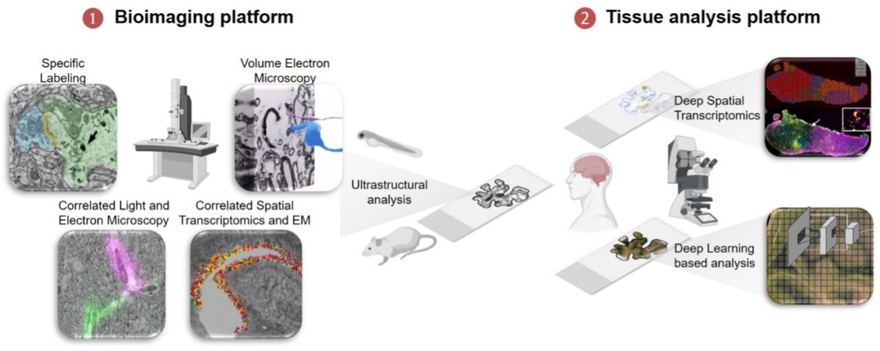
The Z01 Bioimaging and Tissue Analysis Platform supports various CRC projects by analyzing human tissue samples and using state-of-the-art correlative light and electron microscopy techniques to study CNS recovery at a detailed level.
During the first funding period, Z01 contributed to understanding key aspects of CNS recovery, such as axon damage, inflammation’s role in tissue healing, and how certain cells help repair nerve fibers. In the next funding phase, our focus will be on two main goals:
Bioimaging Platform
Develop and refine techniques that combine light and electron microscopy to study how cells in the CNS regenerate. We will enhance ATUM-based correlative light and electron microscopy to observe cellular interactions during CNS recovery with high precision. Additionally, we’ll work on integrating electron microscopy with molecular “-omics” analysis to better understand cellular diversity in damaged or healing tissues, including human samples.
Tissue Analysis Platform
Bridge basic research with human tissue studies by translating experimental findings to human brain tissue. This will be done using a shared microscopy platform that allows researchers at different sites to collaborate and analyze data together. We’ll also continue improving techniques for preserving and analyzing human brain tissue, focusing on integrating cellular structure with gene expression through AI-supported analysis.
Want to collaborate?
Find detailed and relevant information on our password-protected internal page.